Accurate 3D imaging for Covid-19 diagnosis
Professors from LSU Health New Orleans School of Medicine have created a method of Covid-19 diagnosis that uses 3D imaging and modeling techniques that can reliably show the exact spreading path of the virus in human lungs.
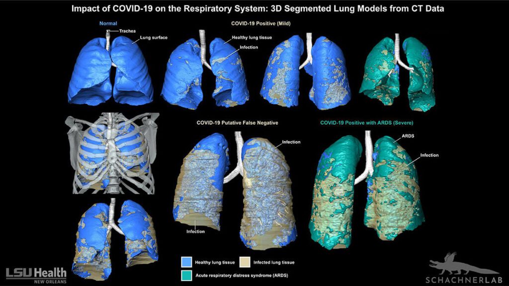
In their paper published in BMJ Case, the researchers describe how the method allows doctors to monitor and path airflow patterns, and lung tissue can be measured volumetrically. This data can be used to make a full 3D model, gives a clearer visualization of the spread and progression of the disease.
The data collected from the scanned patients’ lungs is used to form a map of the lungs that correlates with the virus’ spread and severity. This can aid doctors in quickly distinguishing between other lung pathologies, and COVID-19.
“The full effect of COVID-19 on the respiratory system remains unknown, but the 3D digital segmented models provide clinicians a new tool to evaluate the extent and distribution of the disease in one encapsulated view,” says Dr. Bradley Spieler.
Bradley Spieler is the Vice Chairman of Radiology Research and Associate Professor of Radiology, Internal Medicine, Urology, & Cell Biology and Anatomy at LSU. The techniques used in the 3D Covid-19 diagnosis are those same techniques used for animal anatomy research.
Aside from taking the pressure off hospitals, this Covid-19 diagnosis method can be a fast, accurate, and easily deployable testing technique crucial to any country that wants to ease lockdowns.
“Previously published 3D models of lungs with COVID-19 have been created using automated volume rendering techniques,” says Associate Professor of Cell Biology & Anatomy, Dr. Emma R. Schachner, referring to methods such as volume-rendered models and 2D screen shots of CT scans and radiographs.
With such technologies, hospitals can focus on the extreme cases and utilize resources where necessary. Researchers can now have a clear visual representation of where the virus goes and what it does in the human body.